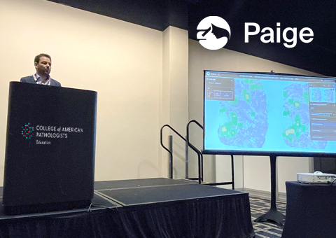
Paige’s Medical Director, Dr. Juan Retamero hosted a product showcase at the 2022 College of American Pathologists (CAP) Annual Meeting where he discussed how artificial intelligence (AI) could be used to help pathologists review breast cases.
Dr. Retamero began with a historical review of pivotal foundational studies that proved that AI was capable of successfully aiding in the identification of cancer with clinical-grade accuracy. One study in particular, Campanella et al., found that multiple instance learning (MIL) was an especially effective model for training the AI 1 such that it could find cancers across data from any institution, regardless of pre-analytical variations, without the need for additional tuning, which is essential for utilizing AI in real-world labs. With this study as a basis, many AI applications have now been brought to the market, including Paige’s, that use this approach to safely identify cancer across prostate, breast, and other specific use cases.
With that context in place, Dr. Retamero then emphasized some of the challenges that breast cancer diagnosis can present, and how AI designed to aid pathologists in breast cancer diagnosis can help to mitigate some of these challenges. He pointed first to a 2015 study that found that breast cancer diagnostic concordance among pathologists against a ground truth diagnosis is not always perfect – in several instances, up to a quarter of pathologists called a benign slide cancerous, and contrarily, several pathologists called a malignant slide benign.2 So, utilizing AI that has been proven to offer high sensitivity for detecting breast cancers, such as Paige Breast, as a second opinion tool could offer enhanced decision support that would help pathologists ensure they are reaching a diagnosis they feel confident in.
A second challenge in breast cancer diagnosis is that the identification of lymph node metastases is often done by a general pathologist rather than a specialist. A study conducted in the Netherlands in 2012 found that when a group of specialists reviewed nodal stages that were diagnosed by general pathologists, up to 24% of these patients had to be upstaged.3 Therefore, AI that has been trained on a robust dataset might offer enhanced sensitivity that could assist all pathologists in diagnosing at a specialist level. Paige breast Lymph Node, in particular, has proven to enhance pathologist efficiency and improve sensitivity in detecting metastases, especially across small micromets that may otherwise be more challenging.4
Additionally, mitotic counting, especially on a microscope, is known among pathologists to be one of the more tedious – if not unlikeable – elements of the diagnostic process. When done manually, it can be hard not only to keep track of the count in a specific region, but to keep track of which regions have already been reviewed and accounted for. This is where AI steps in. AI such as Paige Mitosis Detect can point pathologists to regions with the greatest concentrations of mitosis and automatically calculate the number of mitoses per square millimeter in these hotspots so as to reduce the tedium of mitotic counting and allow pathologists greater diagnostic confidence.
The final stage of diagnosing breast cancers is biomarker assessment across Ki-67, estrogen receptors (ER), progesterone receptors (PR), and HER2, which today is done using immunohistochemistry (IHC), In Situ Hybridization (ISH/FISH), and pathologist assessment alone. However, Dr. Retamero pointed to a paper from 2016 found that when assisted by AI, pathologists showed better differentiation between luminal A and luminal B classification, as well as stronger HER2 classification.5 This suggests that again, as is the case in many other diagnostic processes, the power of pathologists together with AI is stronger than either one alone.
Perhaps the most exciting advancement when applying AI to breast cancer diagnosis though is its potential to change how we approach and diagnose HER2. Traditionally, we have treated HER2 in about 15% of patients.6 Recently, though, it has been shown that a new subset of patients who we consider HER2-low might also benefit from HER2 treatment. The problem is that the HER2 assays we use today are not geared toward diagnosing those patients on the lower end of the HER2 spectrum. AI can change that.
Paige’s AI-powered HER2 assay, HER2Complete, leverages cyto-architectural patterns in tissue to identify subsets of HER2 expression within IHC-0, particularly true HER2-negative disease. Trained via orthogonal proteomic and gene expression methods, HER2Complete can identify HER2 expression in patients currently classified as IHC negative (or IHC-0), in addition to expression in HER2-low (IHC1+ and 2+/FISH negative) patients. This approach is intended to complement existing IHC testing, potentially identifying true HER2-expressing breast cancers using only the diagnostic biopsy or resection slides. Armed with AI, we believe there could be a future where pathologists can identify more patients who might benefit from HER2 treatments.
Dr. Retamero concluded by reminding us that AI tools are designed to support pathologists in reaching a diagnosis, not to diagnose in place of a pathologist. The beauty of AI is only that it creates efficiencies in the work pathologists are already good at and offers additional support in areas that may be challenging. Noting, at the end of the day, it all goes back to morphology – no matter how we slice and dice it. AI is just the means to more effectively tapping into the information that tissues hold. And, armed with this information, a means for helping pathologists guide the entire breast cancer care team with confidence to ensure the best experience for patients.
References
1Campanella, G., Hanna, M.G., Geneslaw, L. et al. Clinical-grade computational pathology using weakly supervised deep learning on whole slide images. Nat Med 25, 1301–1309 (2019).
2Elmore, Joann G., et al. “Diagnostic concordance among pathologists interpreting breast biopsy specimens.” Jama 313.11 (2015): 1122-1132.
3Vestjens JHMJ, Pepels MJ, de Boer M, et al. Relevant impact of central pathology review on nodal classification in individual breast cancer patients. Ann Oncol. 2012;23(10):2561-2566. doi:10.1093/annonc/mds072
4Based on an investigational clinical study involving 3 pathologists and data from 148 patients.
5Stålhammar G, Fuentes Martinez N, Lippert M, et al. Digital image analysis outperforms manual biomarker assessment in breast cancer. Mod Pathol. 2016;29(4):318-329. doi:10.1038/modpathol.2016.34
6Tarantino, P. et al., (2020) J Clin Oncol. 10;38(17):1951-1962.