It has long been recognized among practicing pathologists and within pathology research that breast cancer diagnosis comes with many challenges, including laborious manual processes and diagnostic subjectivity. In recent years, artificial intelligence (AI) designed to resolve those challenges has begun to hit the market. Yet not all pathology AI is created equal. At the Digital Pathology & AI Congress in New York City this month, Dr. David Klimstra, founder and Chief Medical Officer at Paige, explained the key features that make AI lab-safe and why Paige is uniquely positioned to support pathologists in overcoming the hardships of breast cancer diagnosis.
To begin, Dr. Klimstra explained that one of the key factors that sets Paige apart and makes our AI clinic-ready is our innovative approach to model building. We leverage Multiple Instance Learning (MIL), an advanced technique that requires minimal supervision and therefore allows us to train the model on extremely large datasets. In our case, the data comes from Memorial Sloan Kettering Cancer Center, whose world-leading pathologists’ set the gold-standard in diagnosis that is then used as Paige’s ground truth. Importantly, the data was collected from over 200 global institutions to not only ensure that it is representative of a wide range of patient scenarios, but also of a wide range of lab preparation techniques including staining differences and other preanalytical variations. In this way, Paige can build applications that deliver accurate outputs at any lab without the need for on-site tuning.
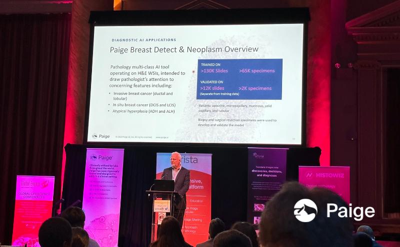
For breast cancer diagnosis, such generalizability is important for overcoming one of the foremost challenges: subjectivity in classification. According to one 2015 study, pathologists had only a 75% overall concordance when diagnosing and classifying breast cancers, and even lower agreement across subtypes like DCIS and atypia. 1 Paige Breast Neoplasm is our application designed to support pathologists in automatically identifying and classifying anything from invasive hyperplasia to invasive carcinoma with exceptional sensitivity and specificity. In doing so without the need for on-site tuning, Paige Breast Neoplasm enables pathologists to reduce diagnostic inconsistency while enhancing diagnostic confidence to ultimately ensure that the patient receives the most accurate possible diagnosis.
The same is true of our AI application for detecting lymph node metastasis, Paige Breast Lymph Node. This diagnostic step is known to be a challenge, especially for generalist pathologists; in one study, up to 24% of patients had their N status upstaged when reviewed by breast experts.2 Paige Breast Lymph Node’s use of robust, gold-standard diagnostic training data allows us to deliver high sensitivity for detecting even small micromets, again across any lab. In this way, it can bridge the gap between specialist and generalist pathologists, minimizing subjectivity and empowering any pathologist to confidently determine N status.
Next, Dr. Klimstra pointed out another unique differentiator of Paige’s AI applications, which is our commitment to the pathologist’s user-experience. Currently, mitotic counting, which is a critical step in diagnosis, is challenging in large part because of the limitations of traditional pathology. On a microscope, the lower power needed to see a concentration of mitoses – otherwise known as a hotspot – is not sufficient to see the mitoses themselves. Instead then, pathologists tend to identify one mitosis and start counting from there. Paige has designed a visualization that allows pathologists to quickly identify a hotspot while still being able to see the individual mitoses, and a grid tool that makes counting those individual mitoses simpler. Paige Breast Mitosis also offers automatic mitotic grades both within that hotspot and for the entire slide, which can greatly reduce the tedium of manual counting. In putting these features in the hands of pathologists, Paige creates seamless ways to enhance efficiency and confidence while allowing pathologists to adapt their workflows to their preferred working style.
It is crucial to note, Dr. Klimstra added, that like the other elements of diagnosis, mitotic counting is an area that is particularly subjective. Paige’s hotspot visualization and other counting functionality make it easy to incorporate AI insights into their diagnosis, but the pathologist will always make the final call. This tool is simply an adjunct, just like immunohistochemistry (IHC) or any other analog diagnostic aid.
Beyond the AI applications themselves, Dr. Klimstra also explained that the Paige Platform’s openness further enhances its value for pathologists and the patients they serve. The vendor-neutral Paige Platform allows seamless integration with laboratory information systems and facilitates access to essential AI tools from third-party vendors like Mindpeak. Without an open platform, this would need to take place in another viewer which is not only cumbersome, but could cause confusion and compromise patient safety should multiple patients be accidentally viewed simultaneously. With Paige’s fully integrated access to Mindpeak’s automated identification and quantification of critical biomarkers such as Ki-67 and HER2, pathologists can streamline workflows, improve efficiency, and reduce subjectivity.
Finally, Paige’s dedication to innovation allows pathologists to adapt to changing patient needs, such as within HER2 grading. Historically, pathologists needed only to classify patients as HER2 amplified or HER2 non-amplified, so all of the existing assays on the market were tuned to support that binary classification. Now, it is important to get more granular in HER2 diagnosis, specifically with regard to HER2-low versus truly HER2 negative. Paige has taken a completely novel approach to HER2 identification, which leverages H&E to identify patients with breast cancer whose tumors have no evidence of expression of HER2. Known as HER2Complete™ BETA, this novel application is intended to complement traditional HER2 testing methods. HER2Complete BETA helps to uncover subsets of HER2 expression within IHC-0 cases, providing clinicians with additional information that could help to inform treatment decisions.
Ultimately, each of Paige’s differentiators contribute to the most important feature of any clinical AI: safety. By building robust, accurate tools, making them simple to use in real-world settings, and innovating new approaches to ongoing challenges, Paige offers a clinic-ready solution that empowers pathologists to make confident decisions and ensure the best possible outcomes for their patients.
To learn more about the Paige Breast Suite, Request a Trial.
—
References
1Elmore JG, Longton GM, Carney PA, et al. Diagnostic Concordance Among Pathologists Interpreting Breast Biopsy Specimens. JAMA. 2015;313(11):1122–1132. doi:10.1001/jama.2015.1405
2Vestjens JHMJ, Pepels MJ, de Boer M, et al. Relevant impact of central pathology review on nodal classification in individual breast cancer patients. Ann Oncol. 2012;23(10):2561-2566. doi:10.1093/annonc/mds072
*In European Union and United Kingdom, Paige Breast Suite AI Applications are CE-IVD & UKCA marked for clinical use with Leica Aperio AT2 and GT450 Scanners. In United States and where research use is permitted, Paige Breast Suite applications use are limited to Research Use Only and not for use in diagnostic procedures.
**HER2Complete BETA™ is released for product evaluation and is currently not available for purchase